Pseudoexfoliation Glaucoma
Pseudoexfoliation glaucoma is type of open angle glaucoma that is caused by small fibrillar, proteinaceous materials.
Pseudoexfoliation is small whitish elastic dandruff like material that is associated with abnormalities in the basement membrane of epithelial cells of the eye such as ciliary epithelial cells, iris pigment epithelial cells and lens capsule epithelial cells.
It can be found in ciliary body, corneal endothelium, zonules, trabecular meshwork, and anterior vitreous but mostly can be seen in the front surface of the lens capsule and also around the pupil margin.
Pseudoexfoliation is also a systemic disease, not only an ocular disease in which this material can also be found in internal organs of the body such as lung, kidney, liver and gallbladder.
This material will flow with aqueous humor and by drained out the eye through trabecular meshwork. This material will accumulate in the trabecular meshwork and with time it will damage the trabecular cells and they will not long be efficient to drain out the aqueous, causing increase in intraocular pressure.
Risk factors of Pseudoexfoliation Glaucoma
1- Race
In is more common Scandinavian countries and rare among African Americans and Eskimos.
2- Sex
Pseudoexfoliation syndrome is more common in females than in males.
3- Age
Pseudoexfoliation syndrome is rarely seen before age 50 years, and its incidence increases steadily with age.
Symptoms and Signs of Pseudoexfoliation Glaucoma
Symptoms of Pseudoexfoliation Glaucoma
1- Patients may be asymptomatic.
2- With chronic disease and with trabecular meshwork damage, high intraocular pressure can lead to pseudoexfoliation glaucoma with blurred of vision and or they may complain of decreased visual acuity secondary to cataract or glaucomatous visual field changes.
Signs of Pseudoexfoliation Glaucoma
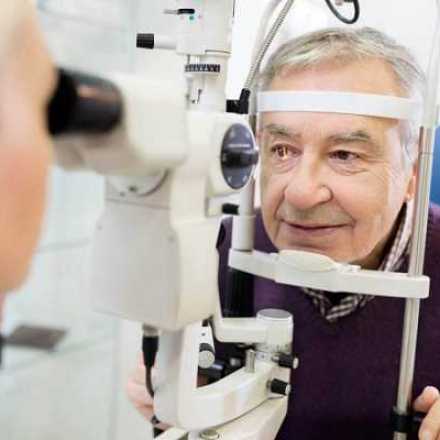
Pseudoexfoliation syndrome can be diagnosed clinically by slit lamp.
1- It is usually bilateral diseases but asymmetrical.
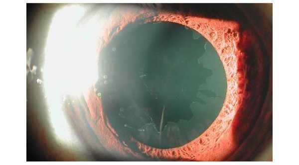
2- Small tiny white material can be seen on the border or margin of the pupil.
3- On the anterior surface of lens capsule. It will form a characteristic ring in this area. This material will form a central disc in the center of the capsule followed be a clear zone which is also followed by a peripheral ring. The central clear zone is due to the movement of the iris over the lens capsule which will brush out the material from this area forming clear zone.
4- In iris transillumination test, there will be patchy atrophy on the papillary margin of the iris.
5- By gonioscopy, there will be increase of trabecular meshwork pigmentation specially in the inferior part of the eye.
6- By gonioscopy, the material can be seen deposited in abnormal location, which is anterior to the trabecular meshwork and this line is called Sampaolesi line.
7- In chronic disease, the IOP can be high when measured by tonometer which can lead to pseudoexfoliation glaucoma.
8- Other signs of glaucoma can be seen such as optic disc cupping, visual field defects.
9- Sometimes, Pseudoexfoliation materials can be deposited in the corneal endothelial cells and can be seen mainly in the inferior half of the cornea.
10- In Pseudoexfoliation, the zonules get weak and in slit lamp we can see that the lens and the iris are not stable and they move. This is called phacodonesis when lens is moving and iridodonesis when the iris is moving.
11- Pseudoexfoliation associated with iris ischemia or reducing blood flow to the iris. This will cause insufficient mydriasis in which the iris will not be fully diluted when mydriatic eye drops applied to the eye
Complications of Pseudoexfoliation Glaucoma
1- Increase in intraocular pressure which with time can lead to secondary open angle glaucoma. Sometimes and because weakness of the zonules, the natural lens will subluxated and move forward to block the pupil. This will cause acute closure angle glaucoma can also occur.
2- Poor iris dilation which is due to iris ischemia. This can be a problem in case of cataract surgery because the iris should be fully dilated to enable the surgeon to remove the cataract without complications. With poor dilated pupil, there will be a high risk of intra-operation complications such as rupture of the posterior capsule and drop of the cataract or part of it to the back of the eye.
3- Weakness of the Zonules. Zonules are special thin fibers that held the natural intraocular lens in place.Due to zonules weakness, there will be phacodonesis which can cause a high incidence of subluxation of lens or even drop of the lens into the posterior part of the eye either during intraocular surgery or after trauma.
Treatments of Pseudoexfoliation Glaucoma
There is a difference between Pseudoexfoliation syndrome and Pseudoexfoliation glaucoma. Not all patients with Pseudoexfoliation syndrome will end up with glaucoma.
There is no treatment for the PEX syndrome but every patient with it should be followed up at least once a year for intraocular pressure measurement.
In case the patient develops pseudoexfoliation glaucoma, the treatment will be with anti glaucoma eye drops and also glaucoma laser surgery in which patient will have excellent respond to laser trabeculoplasty.
In case the patients have poor response to anti glaucoma medications and to laser treatment, surgical treatment is the only option available.


