Pigmentary Dispersion Syndrome
Pigmentary Dispersion Syndrome (PDS) is a benign bilateral condition that characterized by hyperpigmentaion of the trabecular meshwork due to continuous releasing of iris pigments into the anterior chamber.
Etiology of Pigmentary Dispersion Syndrome
There is posterior or back bowing of the iris and with iris movement; there will be continuous rubbing of the back surface of the iris with zonules and the lens. This rubbing will release iris pigments into the aqueous humor in the posterior and the anterior chamber.
Posterior bowing of the iris can be caused by concave shape of the iris and also by reverse papillary block in which the intraocular pressure in the anterior chamber is more than that in the posterior chamber, enhancing backward displacement of the iris. These pigments can deposits in the cornea, trabecular meshwork, lens and also can be seen floated in the aqueous humor.
Risk Factors of Pigmentary Dispersion Syndrome
1- Male sex
2- Young age, 20 to 40
3- Myopic patients
4- Family history
Symptoms of Pigmentary Dispersion Syndrome
It is a symptomatic
Diagnosis of Pigmentary Dispersion Syndrome
It is a symptomatic condition and diagnosed only during the routine ocular examination. We can see these signs during the examination
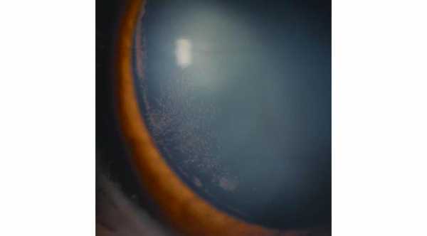
1- Krukenberg's spindle
A vertical band of iris pigments deposition on the inner surface of the cornea| (Endothelial layer), mainly in the lower half of the cornea.
2- By Gonioscopy
Hyperpigmentation of the trabecular meshwork. Normally only the lower part of the trabecular meshwork is hyperpigmented but in PDS, trabecular meshwork is hyperpigmented 370 degree.
3- Iris transillumination
Due to continuous rubbing of the back surface of the iris and the zonules, the iris becomes thin and can transmit lights. Transillumination occurs at the middle part of the iris.
Treatment of Pigmentary Dispersion Syndrome
It is asymptomatic condition that requires no treatment. As patients age, the altered configuration of the cataractous lens may resolve the contact between the iris and the zonules, thus decrease the pigment release.
Although it is a benign condition, patients with PDS should have periodic follow up (3-4 visits/year) with intraocular pressure measurement, Gonioscopy and visual field tests because Pigmentary Dispersion Glaucoma can develop.
Pigmentary Dispersion Glaucoma
It is secondary glaucoma that occurs in patients with Pigmentary Dispersion Syndrome. With time, trabecular meshwork will be blocked and damaged by these iris pigments and intraocular pressure will increase which can cause optic nerve damage if left untreated.
Signs and Symptoms of Pigmentary Dispersion Glaucoma
1- Same signs in patients with PDS plus high intraocular pressure.
2- High intraocular pressure can occur in acute or gradual manner. Acute signs are ocular pain, blurred vision and halos around light.
3- This type of glaucoma can deteriorate rapidly.
Treatment of Pigmentary Dispersion Glaucoma
1- Miotics
Such as Pilocarpine can be used to contract the pupil and decrease contact between iris and zonules and also facilitate the outflow of aqueous humor through trabecular meshwork. It has disadvantages such as exacerbating of myopia which can’t be tolerated by all patients.
2- Eye Drops for Glaucoma
3- Laser Trabeculoplasty https://www.webeyeclinic.com/glaucoma/selective-laser-trabeculoplasty
It can be very effective to these patients.
4- Peripheral Laser Iridotomy
To decrease the posterior bowing of the iris and thus eliminate the contact between the iris and zonules.
5- Trabeculectomy


