

Retinitis Pigmentosa
Retinitis Pigmentosa (RP), also known as pigmentary retinal dystrophy, is a group of inherited diffuse retinal degenerative diseases. It is the most common hereditary dystrophy of the retina. RP is considered as a rare condition.
Definition of Terms:
Retina – the light-sensitive membrane at the back of the eye that contains photoreceptors.
Photoreceptors – are cells in the retina that convert light into electrical signals that are transmitted into the brain and are processed into the images that we see.
Two general types of Photoreceptos:
Rods – located in the outer regions of the retina. Allows us to see in the dark and is responsible for peripheral (side) vision.
Cones – located in the central portion of the retina. Allows us to see colors and fine visual detail.
Symptoms of Retinitis Pigmentosa
Night blindness and dark adaptation difficulties
Loss of peripheral vision (Tunnel vision)
Progressive loss of visual field
Sensitivity to bright lights (Photophobia)
Nearsightedness (Myopia)
Symptoms typically appear in childhood, but progression differs from person to person. However, it is a degenerative condition that eventually leads to significant vision loss or blindness.
In the early stages of retinitis pigmentosa, rods are more affected than cones. As the rods are damaged, the cones eventually breakdown in the late stages of RP leading to more vision loss. Activities of daily living are greatly affected and difficult to perform without assistance.
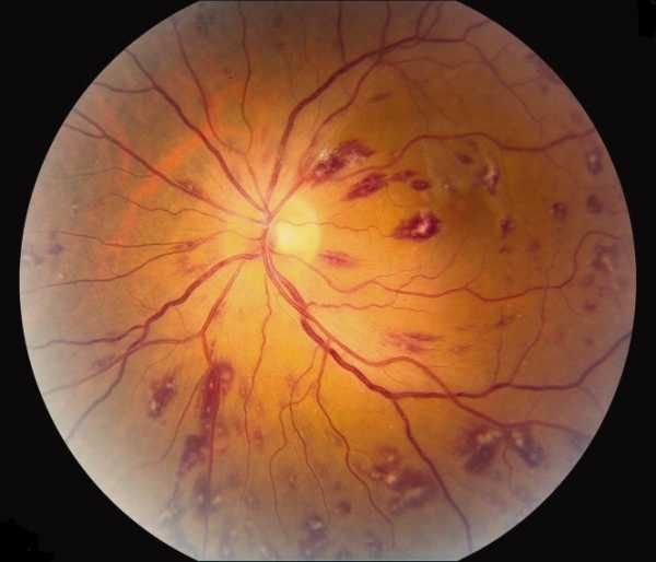
Signs of Retinitis pigmentosa
Pale optic disc and it is called waxy disc pallor
Attenuation of retinal arteries
Pigment Clumpings and dark bone spicule due to hyperpigmentations of retinal pigment epithelial cells
Diagnosis of Retinitis Pigmentosa
Electroretinogram (ERG) – With an ERG, electrodes are placed on and around the eye. A flash of light is sent to the retina and the machine interprets the responses of the rods and cones. Decreased electrical activity from these photoreceptors is indicative of retinitis pigmentosa.
Visual field test – A machine is used to map a person’s field of vision. The patient is asked to fixate on a central light. The patient will be instructed to click a button as they see dots of light emitted by the machine at different locations as they continue to fixate on the central light. The machine then interprets the results and produces a map representing the patient’s visual field.
Genetic testing – A DNA sample is taken from the patient to attain an accurate diagnosis. Genetic testing makes it possible to identify the particular form of the disorder that affects a particular person.
Treatment of Retinitis Pigmentosa
There is no specific treatment available yet, but gene therapy is being studied and seems to be promising. Treatment is mainly supportive.
Low vision aids to maximize remaining vision is beneficial. Different devices are now available, as well as different rehabilitation services to help a person suffering from RP to be able to perform daily activities and somehow maintain their independence.
Some doctors may recommend vitamin A supplementation. Research has shown benefits in slowing the progression of some forms of RP with vitamin A, but monitoring is required as high-doses of vitamin A can be toxic.
Regular follow-up is advised in order to detect any treatable vision-threatening complications.