

Diagnosis of Age Related Macular Degeneration
Often,the diagnosis of Age Related Macular Degeneration is not made until people begin to notice some of the more obvious signs and symptoms of macular degeneration such as blurred vision or distorted vision.
However, ophthalmologists are able to recognize the early stages of AMD even before you begin experiencing any symptoms. So, if you are over the age of 50, you should make regular eye examinations a priority. Early detection of Macular damage or degeneration is an important factor in protecting your eyesight and slowing the progression of the disease.
There are many tests your eye doctor may use to diagnose or to detect early signs of macular degeneration
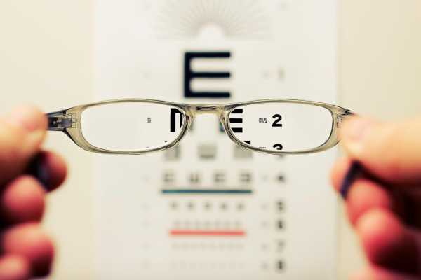
1- Visual Acuity
This test measures the clarity or sharpness of your vision, and will show how well you see objects at various distances. It is usually performed with the same standardized eye chart used to determine eyeglass prescriptions.
2- Amsler grid
This is a very simple test that is used to check macular vision and to detect any damage or disease to the macula, which is located at the center of the retina. Basically, it is a grid that consists of evenly spaced vertical and horizontal lines, resembling a piece of graph paper, with a large dot in the middle.
While staring or focusing at the dot, you will be asked if you see any wavy lines or missing spaces. Any distortion or wavy lines in vision while using amsler grid could indicate the presence of macular damage
3- Tonometry
Using a small instrument called a tonometer, your doctor will measure the intraocular pressure, or the pressure in your eyes. You will be given drops to numb the eyes, making the procedure painless.
You will be given eye drops that will widen or dilate your pupils, allowing the doctor to see the back of your eyes more clearly.
Using a special magnifying lens, your doctor will examine the interior of your eyes, including the retina and optic nerve, looking for signs of damage or disease.
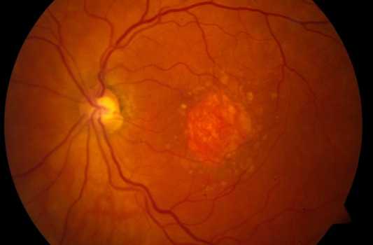
In cases of Age Related Macular Degeneration, typical signs would include white or yellow spots on the retina known as drusens, degeneration of special cells called photoreceptors, and hemorrhaging or scarring within the macula. You may experience blurred vision for a few hours following the dilation of your pupils.
5- Fluorescein Angiography (FFA)
A special dye called fluorescein will be injected into a vein in your arm or hand. This dye will travel through your body, and as the dye enters the vessels in the back of your eye, a special camera will take several photos of your retina. These pictures will help clearly identify any problems that could indicate AMD including the loss or degeneration of cells in the retina, and abnormal or leaking blood vessels.
6- Indocyanine Green Angiography (ICG)
This is similar to FFA with the main difference being the type of dye used. Some doctors believe that ICG is more effective than FFA at identifying some types of eye problems because it offers a deeper examination, allowing ophthalmologists to look underneath the retina for leaking blood vessels. ICG is often used as a secondary test to add more information to results found in the FFA.
7- Optical Coherence Tomography (OCT)
This is a non-invasive test that uses light and light waves to create a high resolution cross-sectional image of the eye's interior. It is very similar to an ultrasound except it uses light rays rather than sound waves to produce the images. Your retina is composed of 10 different layers, and OCT allows each layer to be examined and measured. It will help identify any areas of thickening or thinning within your retina and will allow the doctors see any abnormal masses, veins, or leakage under the retina. OCT is also a helpful follow-up tool after any treatments or procedures, as it can be used to monitor your condition for problems or improvements.
8- Visual Field Testing
This test shows your range of vision, or the horizontal and vertical extent of your field of vision. The doctor will use a series of exercises to check both your central and peripheral vision (side vision), looking for any blind spots or areas of vision loss. Defects in your vision field could have many causes, so your doctor will probably want to do additional testing before making the diagnosis of Age Related Macular Degeneration